


fluorescence imaging is extensively used for visualizing organic tissues including the back of the eye, wherein symptoms of macular degeneration can be detected. it is also usually used to photo blood vessels at some point of reconstructive surgical procedure, allowing surgeons to ensure the vessels are nicely related.
for these procedures, as well as others now in scientific trials, along with imaging tumors, researchers use a part of the light spectrum referred to as the near-infrared (nir) — seven hundred to 900 nanometers, simply beyond what the human eye can come across. a dye that fluoresces at this wavelength is run to the frame or tissue after which imaged the usage of a specialised digicam. researchers have proven that light with wavelengths greater than 1,000 nanometers, called quick-wave infrared (swir), offers lots clearer pictures than nir, however there are no fda-accredited fluorescence dyes with height emission inside the swir range.
a crew of researchers at mit and massachusetts popular medical institution has now taken a first-rate step toward making swir imaging widely available. they have proven that an fda-approved, commercially available dye now used for near-infrared imaging additionally works thoroughly for brief-wave infrared imaging.
“what we located is that this dye, which has been authorised because 1959, is genuinely the nice, the brightest fluorophore that we know of at this point for imaging in the brief-wave infrared,” says moungi bawendi, the lester wolf professor of chemistry at mit. “now clinicians can begin to attempt short-wave imaging for his or her applications due to the fact they have already got a fluorophore which is accepted to be used in people.”
imaging this dye with a digicam that detects quick-wave infrared mild may want to permit docs and researchers to gain lots higher photos of blood vessels and different frame tissues for prognosis and studies.
bawendi and former mit studies scientist oliver bruns are the senior authors of the take a look at, which appears within the proceedings of the countrywide academy of sciences. the paper’s lead authors are mit graduate students jessica carr and daniel franke.
reducing through the fog
the dye that the researchers used in this study, referred to as indocyanine inexperienced (icg), fluoresces most strongly around 800 nanometers, which falls inside the close to-infrared variety. while injected into the frame, it travels via the bloodstream, making it best for angiography (the visualization of blood flowing thru vessels). some robotic-assisted surgical structures have incorporated nir fluorescence imaging to assist visualize blood vessels and other anatomical features.
the mit crew discovered icg’s usefulness for swir imaging fairly serendipitously. as part of a manipulate experiment for another paper, they examined the fluorescence output of quantum dots against the fluorescence output of icg within the short-wave infrared. they predicted that icg would haven't any output, however have been surprised to find out that it certainly produced a totally strong signal.
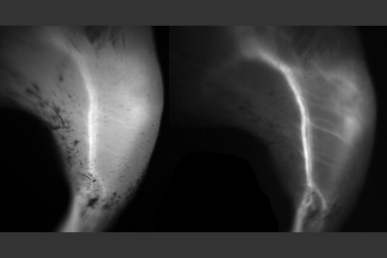
bawendi’s lab and other researchers were interested by growing fluorophores for swir imaging due to the fact swir offers higher contrast and readability than nir. light with shorter wavelengths has a tendency to scatter off of imperfections in items that it moves, but as wavelengths end up longer, scattering is greatly reduced.
“inside the close to-infrared, a whole lot of the capabilities you see in tissue can appearance foggy, and once you circulate into the short-wave infrared, the picture clears up and everything will become sharp,” bruns says.
short-wave infrared also can penetrate deeper into tissue, although calculating precisely how a long way is a complex system, the researchers say, because it relies upon on the size of the shape being regarded and the sector of view of the microscope. inside the new have a look at, the researchers had been able to see several hundred micrometers into tissue using a regular fluorescence microscope. normally, this intensity may be reached most effective with -photon microscopy, a miles more complex and costly sort of imaging.
“we determined that brief-wave infrared is in particular useful for imaging small items that are on pinnacle of a big history, so when you need to do angiography of small vessels, or capillaries, that’s extensively simpler inside the quick-wave infrared than inside the near-infrared,” franke says.
a strong signal
of their take a look at, the researchers similarly explored icg and confirmed that it gives a more potent signal than different swir dyes now in improvement. preceding research of icg had targeted on its emission around 800 nanometers, where it fluoresces the brightest, so no one had observed that the dye additionally produced a sturdy sign at longer wavelengths. even though it doesn’t fluoresce correctly in the shortwave-infrared range, icg absorbs so much mild that if even a small percentage is emitted as fluorescent light, the sign is brighter than that produced through other swir dyes.
the researchers additionally determined that icg is shiny sufficient that it could produce images quick, that is essential for capturing motion.
“if you don’t have a sturdy enough sign, it slows down how lengthy it takes to take the image, so you can’t use it for imaging movement such as blood flowing or the coronary heart beating,” carr says.
the researchers additionally tested any other dye that works within the near-infrared. this dye, called irdye 800cw, is much like icg and can be attached to antibodies that target proteins along with the ones discovered on tumors. they determined that irdye 800cw also fluoresces brightly in the shortwave-infrared mild, concept not as brightly as icg, and confirmed that they could use it to photo a cancerous tumor in the brains of mice.
to do shortwave-infrared imaging, studies labs and hospitals might want to replace from the silicon cameras now used for nir imaging to an indium gallium arsenide (ingaas) digicam. till recently, these cameras were prohibitively high priced, but the fees had been coming down in the beyond several years.
the research crew is now in addition investigating why icg works so nicely for shortwave-infrared imaging, and is trying to perceive the optimal wavelength for its use, which they hope will assist them determine the great applications for this kind of imaging. they're additionally running with different labs to increase dyes which might be just like icg and can paintings even better.
the research become funded by the national institutes of health thru the laser biomedical studies middle; mit through the institute for soldier nanotechnologies; the country wide technological know-how basis; and the department of energy office of science.
0 comments:
Post a Comment