


ucla researchers have produced the clearest three-d pictures to date of the virus that reasons cold sores, herpes simplex virus type 1, or hsv-1. the pix enabled them to map the virus’ structure and supplied new insights into how hsv-1 works.
a document on the research changed into published online by the magazine technological know-how.
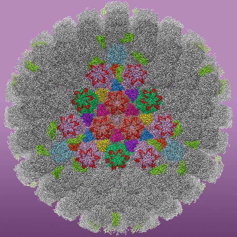
the scientists used cryo electron microscopy, or cryoem, to achieve the primary atomic model of the virus particle, that's made of extra than 3,000 protein molecules comprising tens of tens of millions of atoms.
“we’ve recognized that hsv-1 can disguise in the nucleus of the nerve mobile and set up lifestyles-long latent infection inner most folks,” said xinghong dai, a ucla researcher and the study’s first writer. “but it turned into doubtful how the virus travels from the nucleus of a nerve cellular, alongside the lengthy projection called the axon, and to the pores and skin floor wherein the sore happens. this is the first time we’ve seen how the ones cell shipping motors could bind to their cargo, the hsv-1 capsid ― the protein shell of the virus.”
the observe’s senior creator is z. hong zhou, director of the electron imaging center for nanomachines at the california nanosystems institute at ucla, and a professor of microbiology, immunology and molecular genetics.
till now, scientists have been not able to produce a clean view of the virus to look at its patterns and conduct. zhou and his crew used a way called subparticle refinement to enhance the readability of selected areas of the cryoem images.
the new technique captures designated structural records that could otherwise be difficult to ascertain, in component because the hsv-1 particle is a lot larger than other viruses that cryoem imaging is generally used to look at. this allowed the scientists to provide a specific visualization of the tegument proteins, which seem like five-pointed stars and line the outside of the virus capsid in a normal, or rather ordered, lattice.
“the virus enters and hides inside our neurons in a dormant kingdom, and activates to motive bloodless sores whilst our frame becomes weak,” zhou stated. “these tegument proteins are concerned in transporting the virus inside neurons to our lips to allow lively infection.”
herpes viruses that infect humans are categorized into three subfamilies. in addition to the subfamily that causes cold sores, there may be one that causes start defects and another that reasons most cancers.
the viruses in all three subfamilies percentage similar cores, but range within the protein coat outside the center, the tegument. zhou stated scientists have published papers on the systems of tegument proteins for the beyond 20 years.
“but at decrease resolutions, we didn’t realize precisely what the ones molecules were until now,” he said. “now, we're a hundred percent certain.”
because the capsid-associated tegument complicated, or catc, has precise characteristics compared to those within the different subfamilies, it probably performs a crucial role in hsv-1’s unique existence cycle. with the aid of expertise the steps of this cycle via the structure, zhou and his group received deeper perception into how the virus infects, travels and persists in our nerve cells.
the three-d photograph offers scientists an crucial new tool that could assist factor the way toward the invention and layout of antiviral medications and new remedy alternatives for people with ordinary bloodless sores.
the study became supported in element by means of grants from the national institutes of health and the china scholarship council. contraptions at ucla’s electron imaging middle for nanomachines that had been used to reap images for the published shape are supported by means of ucla and instrumentation presents from the nih and nsf.
0 comments:
Post a Comment